What Happens to Your Tumor Tissue After Surgery
When you're preparing for cancer surgery, your attention is naturally focused on the procedure itself: the surgeon, the anesthesia, the recovery. Very few people stop to ask what happens to the tumor once it's removed. It turns out, most patients have the wrong idea entirely, and that gap has real consequences.
What most patients assume
If you ask most cancer patients what they think happens to their tumor tissue after it's removed, you'll hear one of two answers. Either they assume the hospital keeps it on file permanently, stored somewhere in case it's needed later. Or they assume it's simply returned to them if they ask.
Neither assumption is accurate.
In reality, what happens to your tumor tissue after surgery is determined almost entirely by standard hospital pathology protocol, a process most patients never learn about, and rarely have the chance to influence. By the time you're in recovery, the decision has usually already been made.
Understanding what actually happens is the first step toward making an informed choice.
What actually happens in the hospital
The moment your surgeon removes the tumor, it enters the pathology pipeline. A portion of the tissue is sent to the pathology lab, where technicians examine it under a microscope to confirm the diagnosis, assess margins, and determine staging information.
To do that analysis, the tissue is fixed in a chemical solution called formalin, then embedded in a block of paraffin wax. This process, known as FFPE (formalin-fixed paraffin-embedded), preserves the tissue's structure in a way that allows detailed microscopic examination. It's the standard method used in hospitals worldwide.
Once pathology has what it needs, the remainder of your tumor tissue is typically discarded. There is no universal requirement to store it, and in most institutions, routine surgical specimens are disposed of after the diagnostic work is complete.
A small FFPE block, often just a thin sliver of your original tumor, may be retained in the hospital's archives. How long it's kept depends on the institution: some hold samples for five to ten years, others indefinitely. But storage policies vary widely, and patients are rarely informed of what their specific hospital's policy is.
There is one critical detail in all of this: by the time any of it is archived, the living cells are gone. Formalin fixation kills the cells. What remains is a preserved snapshot of your tumor's structure, useful for certain tests but fundamentally different from living tissue.
Why "dead" vs. "living" tissue matters
FFPE tissue is not without value. It can support standard pathology, immunohistochemistry, and some genomic sequencing tests. For the purposes of diagnosis, it does its job.
But living tissue is a different category of resource entirely.
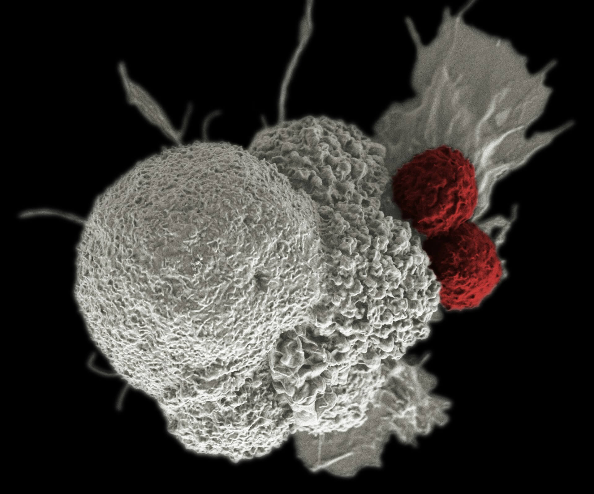
Live tumor cells can be used to grow organoids: three-dimensional miniature tumor models that replicate your specific cancer's behavior outside your body. They can be used for functional drug testing, where researchers can observe how your tumor actually responds to different compounds rather than relying solely on population-level statistics. They are also required for many clinical trial protocols that involve patient-derived tissue.
None of these options are available once the tissue has been fixed in formalin. That process is irreversible. Once your tumor has gone through standard pathology processing, the window for preserving living cells has permanently closed.
This is not a minor technical detail. It's a fundamental fork in the road. What happens to your tumor after surgery determines which future options remain available to you, and which do not.
What you can do instead
You have the right to make decisions about your own biological material. That right extends to your tumor tissue, but exercising it requires acting before surgery, not after.
Custodial biobanking is a service that coordinates the collection and preservation of your tumor tissue at the time of surgery, before it enters standard pathology processing. A portion of the tissue is captured live and preserved across multiple modalities: cryopreserved cells, flash-frozen samples, and FFPE sections. Each preservation method serves different potential uses, and together they give you the broadest possible range of future options.
Timing is everything. The arrangements, including coordination with your surgical team and the pathology lab, have to be in place before the day of surgery. Once the tissue has been removed and processed, there is nothing left to preserve.
If you're planning surgery and want to keep your options open, the conversation needs to happen now.
Bottom line
Your tumor is unique biological information about your cancer. It contains molecular details that no blood test or scan can fully replicate. Under standard hospital protocol, most of that information disappears within days of your surgery, not because it has no value but simply because no one arranged to preserve it.
That doesn't have to be the default. Contacting a custodial biobanking service before your surgery is the one step that keeps those options available. After surgery, the opportunity is gone.
Kernis Health provides concierge coordination and tissue preservation services. This article is informational, not medical advice. Decisions about your care should be made with your oncology team.
Own your biology.
If you or someone you love is preparing for cancer surgery, the best time to plan for tissue preservation is now. Talk to our team and learn what your options are.
Get Started