The Role of Biomarkers in Personalized Cancer Treatment
Cancer treatment has never been uniform, but for much of modern oncology's history, the differences between patients were largely invisible at the molecular level. Two people with the same diagnosis received similar treatment, because there was no reliable way to distinguish their tumors beyond what a pathologist could see under a microscope. That is no longer the case. Today, cancer biomarkers (measurable features of a tumor's biology) give clinicians a way to characterize each tumor individually and use that information to guide care.
A biomarker is any biological indicator that can be measured and tells doctors something clinically useful. In oncology, that might mean how aggressive a cancer is likely to be, how likely it is to recur after initial treatment, or how likely it is to respond to a particular drug. Biomarkers can be proteins expressed on the surface of cancer cells, mutations in the tumor's DNA, patterns of gene activity, or other molecular signals. What they share is that they carry information that, when acted on, can make the difference between a treatment that fits and one that does not.
Understanding what biomarkers are, how they differ, and what it takes to test for them is increasingly part of informed cancer care.
Three Kinds of Cancer Biomarkers
Biomarkers are not a single category. Clinicians and researchers distinguish between types based on what question the biomarker answers, and the distinction matters because different biomarkers are used at different points in care and drive different decisions.
Diagnostic biomarkers help establish whether cancer is present, what kind it is, or where it originated. When a pathologist looks at a tumor sample and runs tests to confirm a diagnosis or identify the primary site of a metastatic cancer, they are often relying on diagnostic biomarkers. These markers help answer the foundational question: what is this?
Prognostic biomarkers provide information about how a cancer is likely to behave over time, independent of any specific treatment. A prognostic marker might indicate that a tumor is likely to grow quickly, is at elevated risk of spreading, or is likely to recur after surgery. This information helps oncologists and patients understand the urgency of the situation and calibrate how aggressively to pursue treatment. Prognostic markers answer the question: what is likely to happen?
Predictive biomarkers are the category most directly tied to treatment decisions. A predictive biomarker indicates whether a particular therapy is likely to work in a particular patient. The presence or absence of a predictive marker tells you something about the relationship between a specific treatment and a specific tumor. Predictive biomarkers answer the question: which treatment is most likely to be effective for this person?
A single biomarker can sometimes serve more than one of these functions. In practice, though, the most clinically consequential development in recent decades has been the expansion of predictive biomarker testing, because it allows treatment to be matched to tumor biology rather than applied uniformly across a diagnosis.
Common Examples in Clinical Practice
Several cancer biomarkers have become standard reference points because of how reliably they guide treatment decisions across large patient populations.
HER2 is a protein that, when overexpressed on breast cancer cells, signals a more aggressive tumor. It is tested routinely in newly diagnosed breast cancer, and the result directly determines whether HER2-targeted therapies are appropriate. HER2 status can also be relevant in gastric and gastroesophageal cancers, making it an example of how a single biomarker can cut across tumor types.
EGFR mutations are among the most well-characterized predictive biomarkers in non-small cell lung cancer. Patients whose lung tumors carry certain EGFR mutations represent a distinct molecular subgroup, one for which targeted therapies have been developed specifically. Testing for EGFR mutations is now standard practice in lung cancer, because the result meaningfully shapes what treatment options are most applicable.
PD-L1 is a protein whose expression level is used to inform decisions around immunotherapy. Tumors that express PD-L1 above certain thresholds have been associated with different patterns of response to immune checkpoint inhibitors. PD-L1 testing is used across a range of solid tumors, including lung, bladder, cervical, and head and neck cancers, and the result is one input, among others, into immunotherapy decision-making.
These three examples represent only a fraction of the biomarkers now in clinical use. Others include BRCA mutations relevant in breast and ovarian cancer, microsatellite instability status relevant across multiple tumor types, and ALK rearrangements in lung cancer, among others. The landscape continues to expand as research matures.
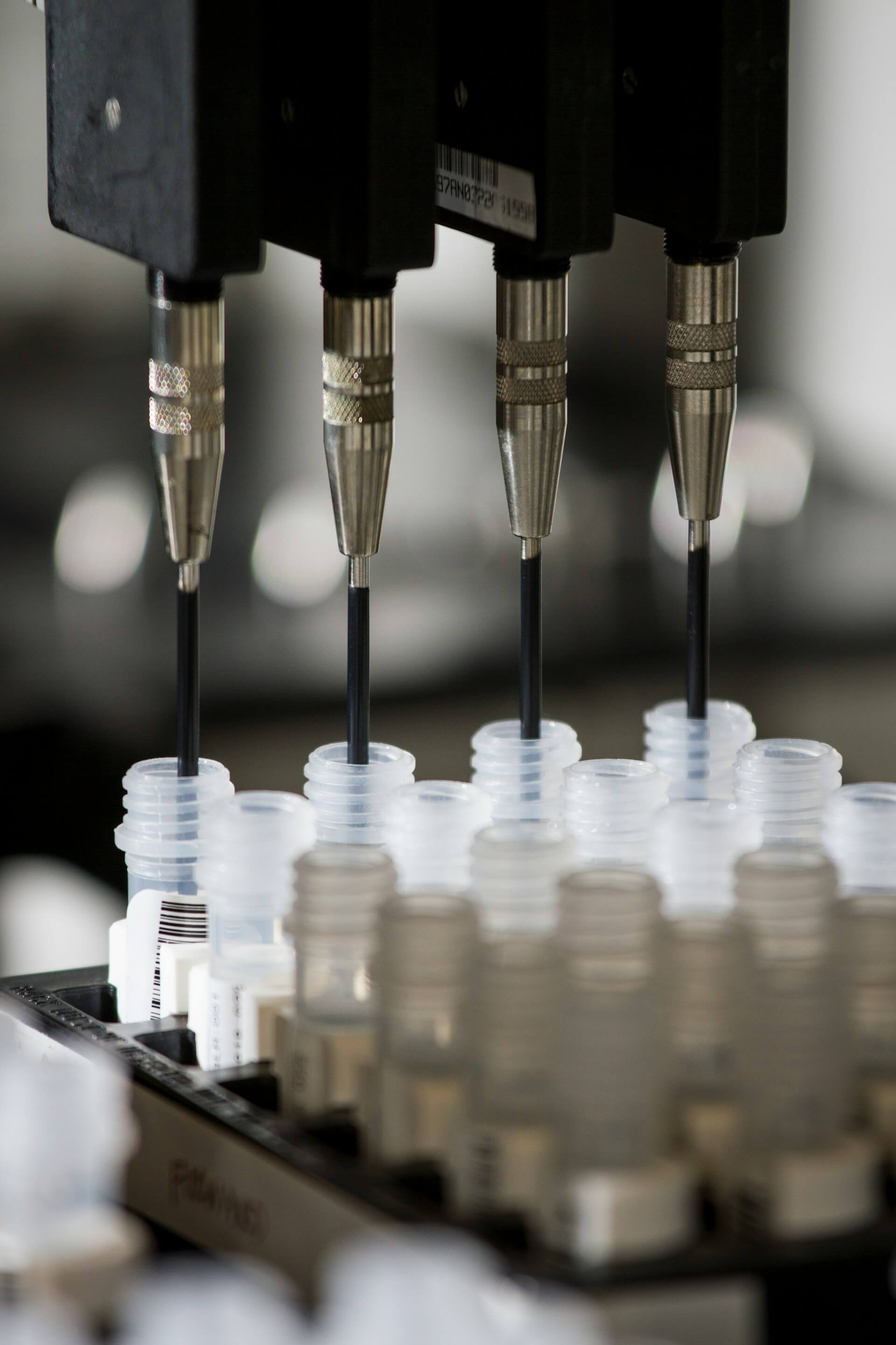
How Biomarkers Are Tested
Biomarker testing is not a single procedure. Different markers require different techniques, and different techniques place different demands on the tissue being tested.
Immunohistochemistry (IHC) is one of the most widely used methods. It applies antibodies to thin sections of tumor tissue to detect the presence and quantity of specific proteins. HER2 and PD-L1 are commonly tested this way. IHC works well with the formalin-fixed, paraffin-embedded (FFPE) tissue that hospitals routinely preserve for pathology.
Genomic sequencing reads the DNA of tumor cells to identify mutations, deletions, amplifications, and other alterations. Targeted panels examine a defined set of clinically relevant genes simultaneously; comprehensive genomic profiling extends this to the full tumor genome. Sequencing can be performed on FFPE tissue, though tissue quality and quantity affect the reliability of results. Liquid biopsy (sequencing cell-free DNA from a blood draw) has emerged as an alternative for some applications when tissue is unavailable, though it has its own limitations.
Gene expression panels measure which genes are active in tumor cells and at what levels. These panels can reveal molecular subtypes that are not visible by diagnosis alone and can help characterize risk or likely behavior. They generally require well-preserved tissue, since the integrity of RNA is particularly sensitive to how tissue is handled after removal.
Choosing the right test, or running multiple tests, depends on the diagnosis and the clinical question being asked. Each test generates information that may not be obtainable through the others.
Why Tissue Type Matters for Biomarker Testing
Not every biomarker test can be performed on every kind of preserved tissue. This is one of the less visible constraints in cancer care, and one that can limit options after the fact if it is not considered in advance.
FFPE tissue, the standard hospital pathology format, supports IHC and many forms of genomic sequencing, making it the most broadly compatible format. But some tests, particularly those requiring intact RNA for gene expression analysis, or those requiring living cells for functional studies, cannot be performed on FFPE material. Fresh or frozen tissue, collected and processed at the time of surgery, is needed for those applications.
This means that the range of biomarker testing available to a patient is partly determined by what tissue was preserved and how. A tumor removed without additional preservation beyond standard pathology may support some testing but not others. Certain questions, including those that might become relevant months or years later as new tests emerge, may simply not be answerable from tissue that no longer exists or was preserved in a format incompatible with the required test.
Bottom Line
Cancer biomarkers are a mechanism for matching treatment to individual tumor biology. Diagnostic biomarkers identify what a cancer is; prognostic biomarkers characterize how it is likely to behave; predictive biomarkers indicate which treatments are most likely to work. Each depends on testing, and each test depends on tissue. Preserving tissue in formats that support the full range of biomarker testing, now and in the future, keeps those options available. The tests that matter most for your care can only be run if the tissue needed to run them has been kept.
Kernis Health provides concierge coordination and tissue preservation services. This article is informational, not medical advice. Decisions about your care should be made with your oncology team.
Own your biology.
If you or someone you love is preparing for cancer surgery, the best time to plan for tissue preservation is now. Talk to our team and learn what your options are.
Get Started