Cryopreservation vs. Flash Freezing vs. FFPE: What Cancer Patients Should Know
Why There Isn't One Method
When people ask about tumor cryopreservation, they are often surprised to learn there are actually three distinct methods for preserving cancer tissue, and none of them is universally superior. The method that is right depends entirely on what the tissue will be used for. Cryopreservation keeps cells alive for dynamic testing. Flash freezing captures a molecular snapshot without preserving viability. FFPE, the format hospitals have used for decades, produces durable slides suited for pathology and standard diagnostics.
Each method opens different doors downstream. Understanding what separates them is not a technical detail. It is fundamental to knowing what your preserved tissue can and cannot do for you.
Cryopreservation: Keeping Cells Alive
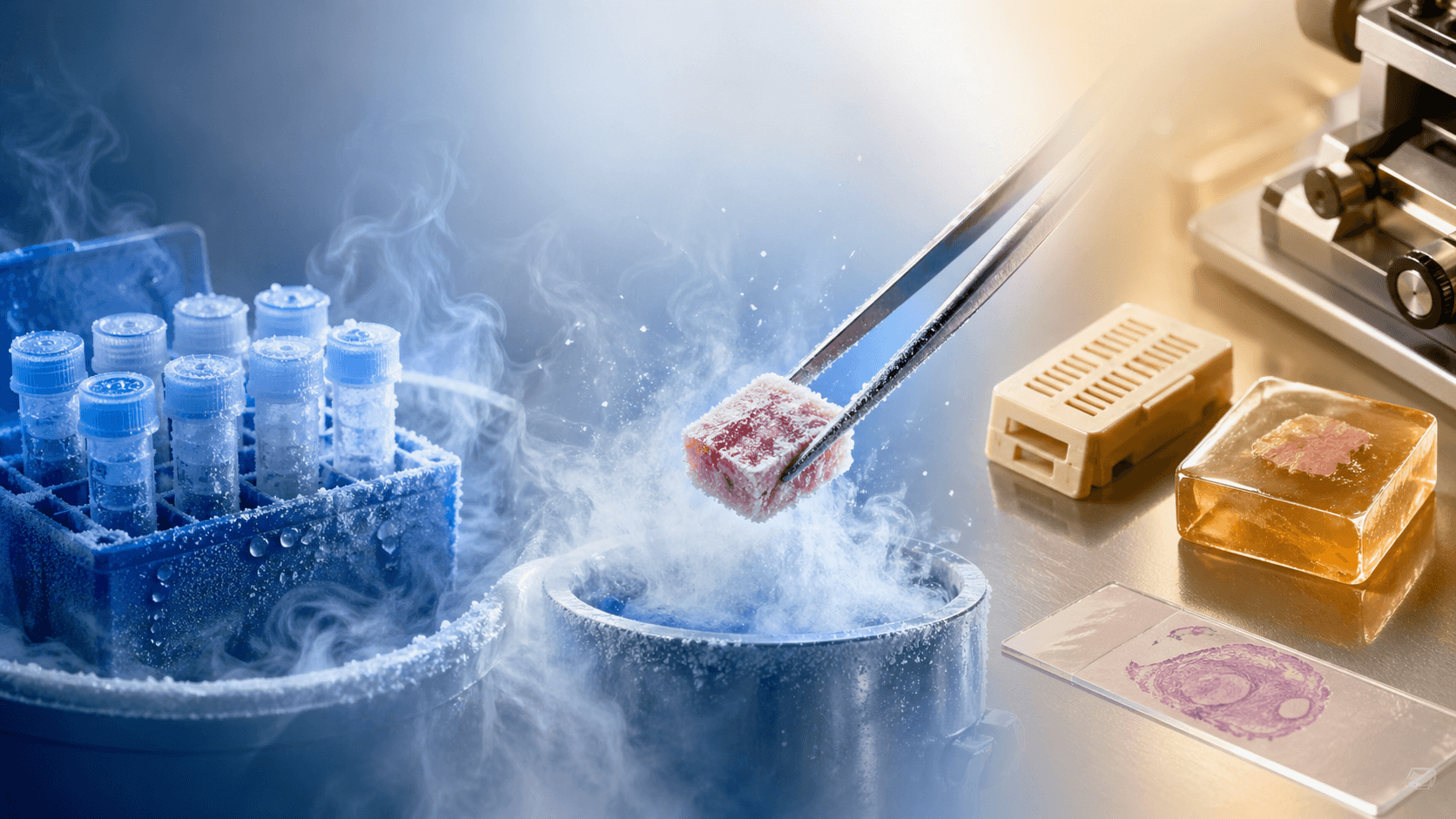
Tumor cryopreservation is the most demanding form of tissue preservation, and the most powerful. The goal is not simply to freeze the tissue. It is to suspend the cells in a state from which they can be fully revived. When done correctly, cryopreserved tumor cells can be thawed weeks, months, or years after collection and resume normal biological activity.
This requires careful handling at every step. The tissue must be transported quickly after resection, processed with cryoprotective agents that prevent ice crystals from destroying cell membranes, and cooled at a controlled rate before being stored at temperatures around minus 196 degrees Celsius in liquid nitrogen. Any deviation (a delay, a temperature fluctuation, improper media) can compromise viability and render the cells useless for live-cell applications.
The payoff for that precision is significant. Cryopreserved tumor cells are the only format that enables organoid development: the growth of three-dimensional, miniature tumor models derived directly from a patient's cancer cells. Organoids replicate the structural and functional characteristics of the original tumor, making them a uniquely patient-specific research tool.
Live-cell drug sensitivity testing also depends on cryopreservation. In this application, thawed tumor cells are exposed directly to a panel of treatment compounds, and their response is measured. This kind of testing requires that the cells be biologically active: they must be capable of growing, dividing, and dying in response to drugs. FFPE and flash-frozen tissue cannot support this.
Many clinical trials and translational research programs also specify cryopreserved tissue as an eligibility requirement. As personalized medicine advances, the demand for viable tumor cells is growing. Tumor cryopreservation is the only method that keeps that door open.
Flash Freezing: A Molecular Snapshot
Flash freezing, also called snap freezing, takes a different approach. Rather than trying to preserve cellular viability, it aims to arrest all biological activity at the moment of collection. Tissue is plunged into liquid nitrogen within seconds of resection, instantly stopping every enzymatic process and locking the molecular contents of the cells in place exactly as they were at that moment.
The cells themselves do not survive. The rapid temperature drop is incompatible with cell viability. But the molecular state of the tissue (its DNA, RNA, proteins, and metabolites) is preserved with high fidelity. This is the format that underpins the most analytically rich studies in cancer research.
Whole-genome sequencing, RNA expression profiling, proteomics, and metabolomics all depend on high-quality, non-degraded molecular material. Flash-frozen samples provide that. The integrity of RNA in particular is highly sensitive to degradation; flash freezing stops the RNases that would otherwise begin breaking it down within minutes of collection.
A flash-frozen archive becomes more valuable over time, not less. New analytical tools continue to emerge: methods that can extract information from preserved tissue that could not have been obtained at the time of collection. A sample frozen today may answer questions that have not yet been asked.
FFPE: The Hospital Standard
Formalin-fixed, paraffin-embedded tissue is what your hospital's pathology department uses, and has used for over a century. After resection, tissue is immersed in formalin, a chemical that cross-links proteins and halts degradation. It is then embedded in paraffin wax, which allows the block to be sliced into thin sections, mounted on glass slides, and examined under a microscope. FFPE blocks are stable at room temperature for years, even decades.
This durability and the extensive infrastructure built around it make FFPE the foundation of clinical pathology. A pathologist examining your tumor to confirm diagnosis, assess surgical margins, and stage your cancer is almost certainly working from FFPE slides. The format is also compatible with immunohistochemistry (staining techniques that reveal the presence and location of specific proteins) and with standard DNA-based tests like next-generation sequencing panels.
But FFPE has meaningful limitations. Formalin fixation chemically alters the tissue in ways that degrade certain molecules, particularly RNA. The cross-linking process that makes the sample stable also introduces artifacts that can confound some modern analyses. FFPE cannot support live-cell applications at all. The fixation process kills every cell.
The hospital keeps FFPE for its own clinical purposes. That material serves an institutional diagnostic function. It is not necessarily stored indefinitely, it may not be accessible to you on request, and it exists in a format that cannot serve many of the most analytically powerful downstream uses. It is a starting point, not a complete answer.
Method Comparison
| Method | What It Preserves | Best For | Limitations |
|---|---|---|---|
| Cryopreservation | Living, viable cells | Organoid models, live-cell drug sensitivity testing, clinical trials requiring viable tissue | Requires precise handling and unbroken cold chain from collection; most demanding to execute |
| Flash Freezing | Molecular contents (DNA, RNA, proteins, metabolites) in high-fidelity state | Genomics, transcriptomics, proteomics, metabolomics, long-term molecular archives | Cells are not viable; requires liquid nitrogen storage; cannot support live-cell applications |
| FFPE | Tissue architecture and protein structure for histological examination | Pathology review, immunohistochemistry, standard next-generation sequencing panels | Cells are not viable; formalin cross-linking degrades RNA; limits advanced molecular analysis |
Why Multi-Modal Preservation Is the Ideal
The strongest position a cancer patient can be in is to have tissue preserved across all three modalities. No single method covers every possible downstream use. An organoid study requires cryopreserved cells. A comprehensive molecular analysis of gene expression requires flash-frozen material. A second pathology review or a standard diagnostic test may require FFPE.
Future treatment options are difficult to predict. New clinical trials open, new testing platforms emerge, and the science of cancer continues to advance. Each of those developments may require a different type of preserved material. A multi-modal biobank, one that holds cryopreserved, flash-frozen, and FFPE samples, provides the broadest possible optionality going forward.
The opportunity to collect tissue across all three formats exists only at the time of surgery. What is not preserved in that window cannot be recovered. Building a comprehensive tissue archive at the outset is how patients keep the most doors open.
Kernis Health provides concierge coordination and tissue preservation services. This article is informational, not medical advice. Decisions about your care should be made with your oncology team.
Own your biology.
If you or someone you love is preparing for cancer surgery, the best time to plan for tissue preservation is now. Talk to our team and learn what your options are.
Get Started